Course Syllabus
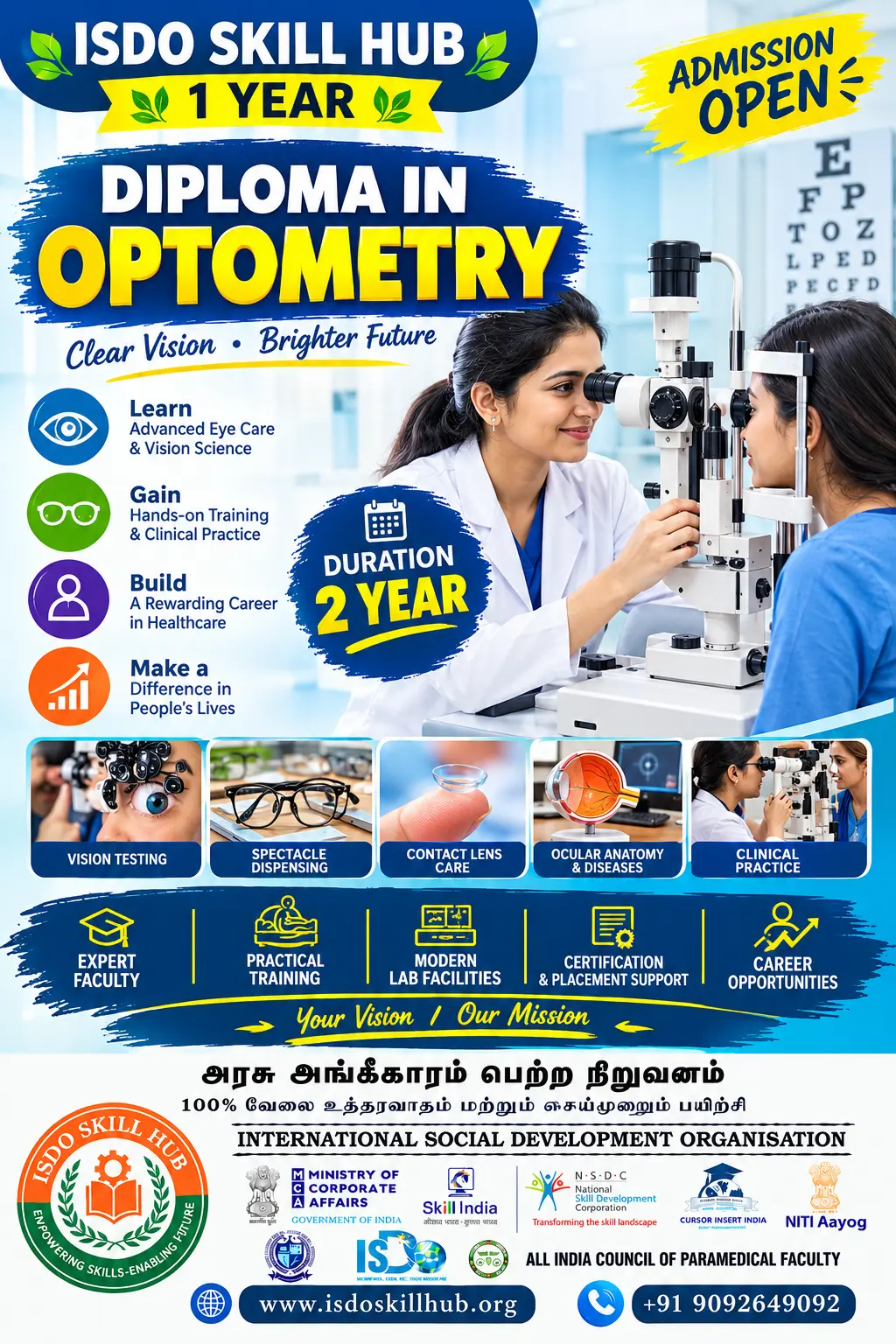
Diploma in Ophthalmic Technology
Duration: 2 Years
Level: Diploma
Units: 10
Mode: Theory + Practical
Unit 1: Fundamentals of Ophthalmology & Ocular Anatomy
Theory
- Introduction to ophthalmology and ophthalmic technology
- Role and responsibilities of an Ophthalmic Technician/Technologist
- Anatomy of the eye and adnexa
- Physiology of vision
- Cornea, conjunctiva, sclera and anterior chamber
- Lens, retina, optic nerve and visual pathway
- Basic ophthalmic terminology
Practical
- Identification of ocular structures using models and charts
- Basic patient communication
- Ophthalmic history-taking under supervision
- Clinical observation and documentation
- Introduction to ophthalmic examination workflow
Unit 2: Visual Acuity & Basic Vision Assessment
Theory
- Principles of visual acuity
- Distance and near vision
- Snellen and LogMAR charts
- Pinhole testing
- Colour vision assessment
- Basic contrast and visual function
- Documentation of visual acuity
Practical
- Distance visual acuity testing
- Near vision testing
- Pinhole testing
- Colour vision testing
- Patient positioning and instructions
Today 10:44 AM
Provide the 2 Year , 10-unit Theory + Practical syllabus, final project, assessment, and course tags
DIPLOMA IN OPHTHALMIC TECHNOLOGY
Duration: 2 Years
Level: Diploma
Units: 10
Mode: Theory + Practical
UNIT 1: Fundamentals of Ophthalmology & Ocular Anatomy
Theory
- Introduction to ophthalmology and ophthalmic technology
- Role and responsibilities of an Ophthalmic Technician/Technologist
- Anatomy of the eye and adnexa
- Physiology of vision
- Cornea, conjunctiva, sclera and anterior chamber
- Lens, retina and optic nerve
- Visual pathway
- Basic ophthalmic terminology
Practical
- Identification of ocular structures using models and charts
- Basic patient communication
- Ophthalmic history-taking under supervision
- Patient identification and documentation
- Introduction to ophthalmic clinical workflow
UNIT 2: Visual Acuity & Basic Vision Assessment
Theory
- Principles of visual acuity
- Distance and near vision
- Snellen and LogMAR charts
- Pinhole testing
- Colour vision
- Contrast sensitivity fundamentals
- Visual-field concepts
- Recording and documentation of visual findings
Practical
- Distance visual-acuity testing
- Near-vision testing
- Pinhole testing
- Colour-vision assessment
- Basic contrast-sensitivity demonstration
- Patient positioning
- Recording visual-acuity results
UNIT 3: Optics & Refraction
Theory
- Fundamentals of light and optics
- Reflection and refraction
- Refractive errors
- Myopia, hypermetropia and astigmatism
- Presbyopia
- Spherical and cylindrical lenses
- Trial-frame principles
- Subjective and objective refraction
- Introduction to retinoscopy
Practical
- Identification of lenses
- Lens-power measurement
- Trial-frame preparation
- Trial-lens arrangement
- Basic subjective-refraction procedures under supervision
- Retinoscopy demonstration
- Recording refractive findings
UNIT 4: Ophthalmic Instruments & Equipment
Theory
- Introduction to ophthalmic instruments
- Slit-lamp biomicroscope
- Direct and indirect ophthalmoscope
- Tonometer
- Keratometer
- Lensometer
- Ophthalmic imaging equipment
- Equipment safety and maintenance
Practical
- Identification of ophthalmic instruments
- Instrument setup
- Slit-lamp examination observation
- Ophthalmoscope handling
- Tonometer demonstration
- Keratometer operation
- Lensometer operation
- Basic cleaning and maintenance
UNIT 5: Ocular Examination & Diagnostic Procedures
Theory
- Basic eye-examination sequence
- External eye examination
- Anterior and posterior segment examination
- Pupil assessment
- Ocular motility and alignment
- Tonometry principles
- Fundus examination
- Basic diagnostic documentation
Practical
- Preliminary eye examination
- Pupil assessment
- Ocular motility observation
- Tonometry under supervision
- Slit-lamp examination
- Fundus examination demonstration
- Recording examination findings
UNIT 6: Ophthalmic Imaging & Specialized Investigations
Theory
- Principles of ophthalmic imaging
- Fundus photography
- Optical Coherence Tomography (OCT)
- Visual-field testing
- Corneal topography
- Keratometry
- Ocular biometry
- Image quality and documentation
Practical
- Fundus-camera setup demonstration
- Fundus image acquisition under supervision
- OCT procedure observation
- Visual-field testing
- Keratometry practice
- Corneal-topography demonstration
- Basic ocular-biometry workflow
- Image-quality assessment
UNIT 7: Common Eye Conditions & Clinical Support
Theory
- Refractive errors
- Cataract
- Glaucoma
- Conjunctivitis
- Corneal disorders
- Retinal disorders
- Diabetic eye disease awareness
- Common pediatric eye conditions
- Age-related eye conditions
- Warning signs requiring clinical referral
Practical
- Patient history documentation
- Vision assessment for different age groups
- Preparation for ophthalmic examinations
- Observation of common clinical findings
- Patient education on routine eye care
- Referral and escalation documentation
UNIT 8: Ophthalmic Surgery, Sterilization & Patient Care
Theory
- Introduction to ophthalmic surgery
- Common ophthalmic surgical procedures
- Pre-operative patient preparation
- Ophthalmic surgical instruments
- Sterilization and disinfection
- Infection prevention and control
- Post-operative patient support
- Eye-care safety and emergency awareness
Practical
- Pre-operative checklist preparation
- Identification of ophthalmic surgical instruments
- Surgical tray arrangement
- Sterile-field awareness
- Hand hygiene and PPE
- Instrument cleaning and sterilization workflow
- Post-operative patient-care observation
- Surgical documentation
UNIT 9: Contact Lens, Low Vision & Rehabilitation
Theory
- Introduction to contact lenses
- Types and basic applications of contact lenses
- Contact-lens care and hygiene
- Introduction to low vision
- Functional vision assessment
- Low-vision aids
- Visual rehabilitation principles
- Patient counselling and education
Practical
- Contact-lens identification
- Contact-lens care demonstration
- Hygiene and handling procedures
- Basic low-vision assessment
- Identification of low-vision aids
- Demonstration of assistive devices
- Patient education and counselling
UNIT 10: Integrated Ophthalmic Technology Practicum
Theory
- Complete ophthalmic examination workflow
- Patient preparation and communication
- Visual-acuity assessment
- Basic refraction workflow
- Ophthalmic diagnostic procedures
- Imaging and equipment management
- Infection control
- Documentation and quality assurance
- Professional ethics and career development
Practical
- Complete preliminary eye examination
- Visual-acuity testing
- Basic refraction under supervision
- Slit-lamp examination
- Tonometry
- Fundus examination
- Ophthalmic imaging procedures
- Patient preparation
- Clinical documentation
- Equipment maintenance
- Supervised clinical ophthalmic practicum
FINAL PROJECT
Comprehensive Eye Examination & Ophthalmic Diagnostic Project
Students will complete a supervised project demonstrating the workflow of a selected ophthalmic assessment.
Project Components
- Selection of an approved clinical case
- Patient identification and history
- Visual-acuity assessment
- Basic refractive assessment
- Ocular examination under supervision
- Appropriate ophthalmic instrument selection
- Diagnostic-test workflow
- Ophthalmic imaging where applicable
- Recording and documentation of findings
- Basic quality-control checklist
- Patient education and referral awareness
- Final case report and presentation
Project Deliverables
- Project proposal
- Patient-assessment form
- Visual-acuity record
- Examination checklist
- Sample/approved diagnostic images
- Equipment checklist
- Clinical logbook
- Final project report
- Practical demonstration
- Viva and presentation
Important: Diagnostic interpretation, prescribing, surgical procedures and other restricted clinical activities must be performed by appropriately qualified professionals. Student practical work should remain within the scope permitted by the institution and applicable regulations.

 Bestseller